


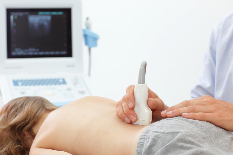
Ultrasonography is the practice of being able to visualise areas inside the body by using ultrasonic pulses. This is of course great for various medical purposes, such as those that fall into the category of orthopedics.
Ultrasonography and Orthopedic Diagnosis
Some healthcare professionals are of the opinion that ultrasonography isn’t of use within the field of orthopedics because ultrasounds can not penetrate bones. However, these machines can be used to diagnose fractures and other similar health problems.
Ultrasounds have also been known to be used within surgical settings. When used properly, an ultrasonography machine is an incredibly versatile tool that can be of great benefit during the diagnostic process.
Bones and Ultrasounds
Bones are able to reflect the waves from the ultrasound because of their structure and density. With this lack of signal nothing is visible beyond the cortical surface besides acoustic shadows. There is no diagnostic benefit to being able to see the shadows.
Although there is not a great deal to be seen when assessing bones with an ultrasonography machine, this certainly does not rule it out as a great diagnostic tool. Valuable information can still be determined from the reflection of the surface of the bone in question. It is particularly useful at diagnosing tumours on the bone as the blood flow to the tumour can be assessed.
Ultrasonography and Tumour Diagnosis
When it comes to tumour diagnosis, the ultrasonography is of great use as it can show the doctor exactly where in the body the tumour is located. This is then useful for the process of performing a biopsy. An ultrasound scan will not be able to determine if a tumour is cancerous. It simply serves as a way to ascertain the specific location of the tumour in question.
This kind of diagnostic procedure may be suggested if an initial X-ray isn’t adequate to present the tumour to the healthcare professional. If the desired results are still not achieved than a CT scan or an MRI will generally be able to provide greater detail.
Preparing for an Ultrasound
If your healthcare professional advises you that you will need an ultrasound then you will be given specific instructions beforehand. These instructions will generally vary depending on the part of the body that is being investigated. However, there are certain things that are always best practice, such as not eating for several hours before your scan takes place. For scans involving the pelvic area it is usually recommended that you drink water an hour before your scan and avoid using the toilet until afterwards.
In certain cases a sedative will be prescribed to relax you before your ultrasound. However, if this is not the case then you will be able to return home immediately after your scan. There are no after-effects or side-effects of the ultrasound itself. If you were given an ultrasound and you do not have someone to accompany you home, then you will have to stay in the hospital until the medication wears off.
Having an ultrasound is generally a painless process, and at most you may experience some slight discomfort. One thing to be aware of is that a latex cover may be used on the probe. So, if you are allergic to latex then you should make the sonographer aware of this as soon as possible.
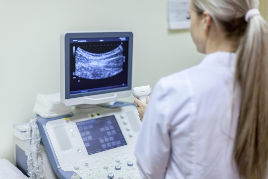
The Ultrasonography Machine
When an ultrasonography machine is used, it sends the ultrasonic pulses directly into the body tissue. The frequency used during this process will generally range from 1.6 megahertz up to 10 megahertz.
As with all things, there are certain advantages and disadvantages to using the ultrasonography machine. It is by no means the only imaging device that can be used when diagnosing a problem. For example, some orthopedic practices may consider it too expensive to do an ultrasound.
Traditionally ultrasounds have been used to assess unborn babies, breasts, blood vessels, hearts and glands, as well as the organs within the abdomen. However, using ultrasounds to evaluate the musculoskeletal system is now beginning to increase in popularity and prominence. It is now more common for those studying healthcare to be taught how to read a musculoskeletal sonogram. This means that it will continue to be used more as those entering the field of radiology will already have it as a skill and know of its benefits.
At Onto Orthopedics we are excited to be able to offer this diagnostic method to our patients. We believe it enables us to offer a superior service and reach the correct diagnosis as quickly as possible!
References
1) http://study.com/academy/lesson/what-is-ultrasonography-definition-history-uses.html
2) http://www.nhs.uk/conditions/ultrasound-scan/Pages/Introduction.aspx
Related Posts


Cigarettes May Inhibit Inflammation Treatments
Axial spondyloarthritis, also known as AxSpa, is a chronic…